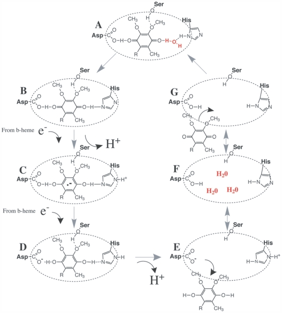
Figure 6. The reduction of ubiquinone to ubiquinol in the Qi binding site in cytochrome b.
(A) Shows ubiquinone bound in the Qi pocket to surrounding cytb residues. A water molecule involved in H-bonding is highlighted in red. The reduction of ubiquinone proceeds from (B) to (D) with the vacation of ubiquinol shown in (E). (F) Shows the vacant Qi site with replenished water molecules (red) that replace the H+ used in the reaction. The cycle continues in (G) with another ubiquinone entering the binding site and the process starts again. The figure was adapted from Kolling et al. [38] and Crofts [36].

