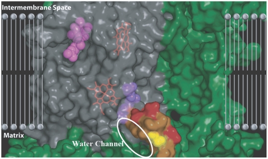
Figure 7. The water channel that leads up into the hydrophobic membrane region where the Qi binding site is embedded.
The approximate location of the membrane was obtained from Gao et al. [35]. The electrostatic surface is shown for the residues of complex III (only one monomer shown). Cytb is colored in grey and the surrounding subunits are shown in green. The CoQ coenzymes (pink and purple) are shown bound in their respective sites. The N-terminus tail of cytb has been highlighted according to the hydropathy of the residues in order to show the potential path for water to be shuttled up into the Qi site. Hydrophilic residues on the cytb tail are highlighted in orange and hydrophobic residues are highlighted in red. The figure shows a potential hydrophilic channel up the cytb tail that allows water into hydrophobic membrane and into the Qi site. The cytbI7T polymorphism (yellow) resides in the center of this water channel.

