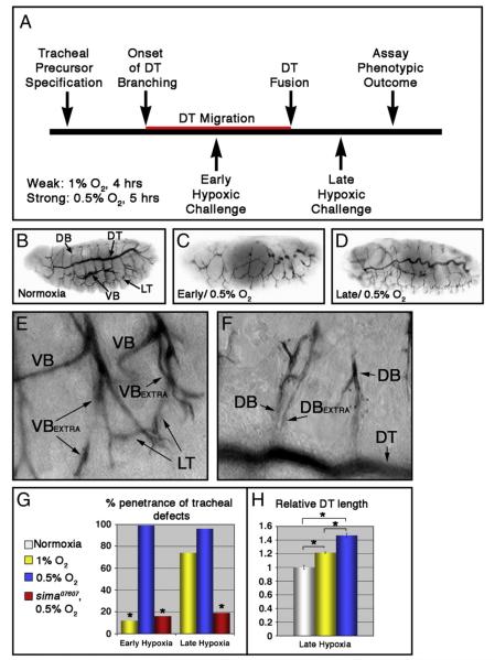
Fig. 1.
Tracheal morphogenesis defects following exposure to hypoxia. (A) Schematic representation of severity and timing of hypoxic challenges, relative to migration of dorsal trunk (DT) fusion cells (in red). (B—F) Lateral images of w1118 embryos stained with the tracheal lumen marker mAb2A12. Unless otherwise indicated, embryos are shown anterior left, and dorsal up. (B) Tracheal architecture of a normoxic embryo. The DT, dorsal branches (DBs), lateral trunk (LT) and visceral branches (VBs) are indicated. (C—F) Hypoxia treated embryos showing characteristic phenotypes following exposure to 0.5% O2. (C) Early hypoxic exposure leads to defects in tracheal morphogenesis. (D) Sinuous overgrowth seen following late hypoxic exposure. (E, F) Late hypoxic exposure also causes duplications of the (E) VB and (F) DB within given segment. ‘Extra’ branches are indicated. (G) Quantification of penetrance of tracheal defects in the indicated hypoxic conditions and genotypes (*p<0.001 relative to w1118 embryos in 0.5% O2 [blue bars]). (H) Quantification of DT length in the indicated hypoxic conditions relative to normoxic control, showing a graded hypoxic response (*p<0.005; error bars are ±standard error of the mean [SEM]).

