Fig. 2.
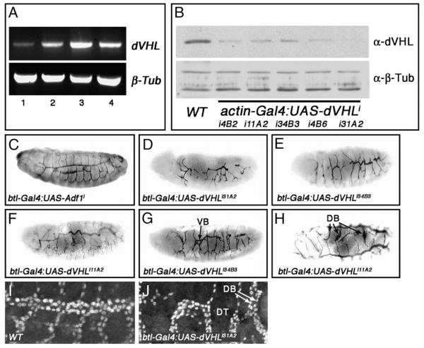
Tracheal-specific knockdown of dVHL leads to defects in embryonic tracheal morphogenesis. (A) RT-PCR analysis of dVHL (top) and β-tubulin (bottom) expression during embryonic stages 1–8 (lane 1), 9–11 (lane 2), 12–14 (lane 3) and 15–17 (lane 4). (B) Western blot analysis of dVHL (top panel) levels in whole embryo extracts from stage 13–16 control embryos (wt) and embryos expressing the indicated combination of actin-Gal4 and UAS-dVHLi lines. α-β-Tubulin is used a loading control (bottom panel). (C—G) Lateral and (H) dorsal images of embryos stained with the tracheal lumen marker mAb2A12. (C) Normal tracheal architecture in a btl-Gal4-UAS-Adf1 control RNAi embryo (D—F) Embryos of the indicated genotypes showing the range of tracheal defects seen following btl-Gal4:UAS-dVHLi knockdown. (G,H) btl-Gal4 driven dVHL knockdown also causes duplications of (G) VBs and (H) DBs as indicated. (I, J) Lateral images of (I) w1118 and (I) btl-Gal4:UAS-dVHLi31A2 embryos stained with α-Tgo to mark tracheal cells. (J) Magnified view of a dVHLi embryonic trachea showing DT interruption, and missing (asterisk) or duplicated (arrows) DBs.

