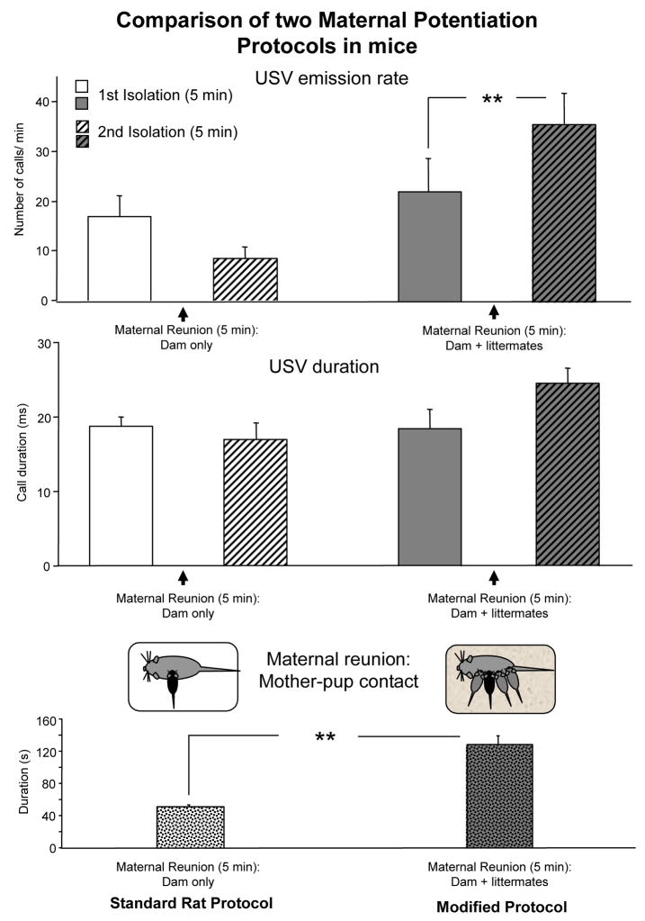
Figure 2.
Effects of two maternal potentiation protocols in 8-day-old C57Bl/6J mice: in the first method, shown in the left column, maternal reunion between first and second isolation consisted in returning the pup to dam only, following the standard protocol for rats. In the second protocol, shown in the right column, maternal reunion consisted of returning the pup to the home cage with the dam and the littermates. Data are mean + sem, n = 10 litters per protocol [methods for the USV recording and statistical analysis are as described in Scattoni et al. 2007]. Upper panel: number of USV per min emitted by the experimental pup (black pup in the diagram) during the first and second isolation periods, ** p< 0.01 after Tukey HSD posthoc comparison performed on the interaction protocol x maternal potentiation factor F (1, 18) =23.6, p<0.01. Middle panel: mean duration of USV emitted by the experimental pup (black pup in the diagram) during the first and second isolation periods; protocol x maternal potentiation factor F (1, 18) =4.5, p<0.05, Tukey HSD posthoc comparison performed on this interaction just missed statistical significance. Lower panel: time spent by the dam in active contact with the experimental pup (black pup in the diagram) during maternal reunion, ** main effect of protocol F (1, 18) =48.9, p<0.01.

