
Figure 2. SRp38(−/−) mice have multiple heart defects and severe liver damage.
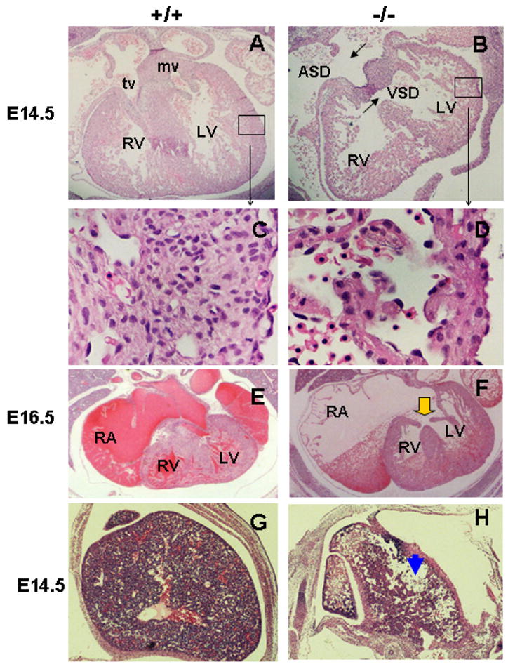
HE staining was performed on heart transverse sections of SRp38 wild-type (A,C,E) and mutant (B,D,F) embryos to examine cardiac morphology at E14.5 (A–D) and E16.5 (E–F). SRp38(−/−) hearts have atrial septal defects (ASD) and ventricular septal defects (VSD) at E14.5 (compare A and B). High-power view of the left ventricle (boxed area) shows disorganized cardiomyocytes and thin myocardium in the mutant as compared to the wild-type littermate (C). Atrioventricular canal defect (AVCD, arrow) and enlarged right atria (RA) are observed in the SRp38(−/−) heart at E16.5 (compare E and F). RV, right ventricle; LV, left ventricle; tv, tricuspid valve; mv, mitral valve. HE-stained transverse sections of the liver showed large amounts of tissue loss in the SRp38(−/−) embryos (H, arrowhead, compared to wild-type control G)