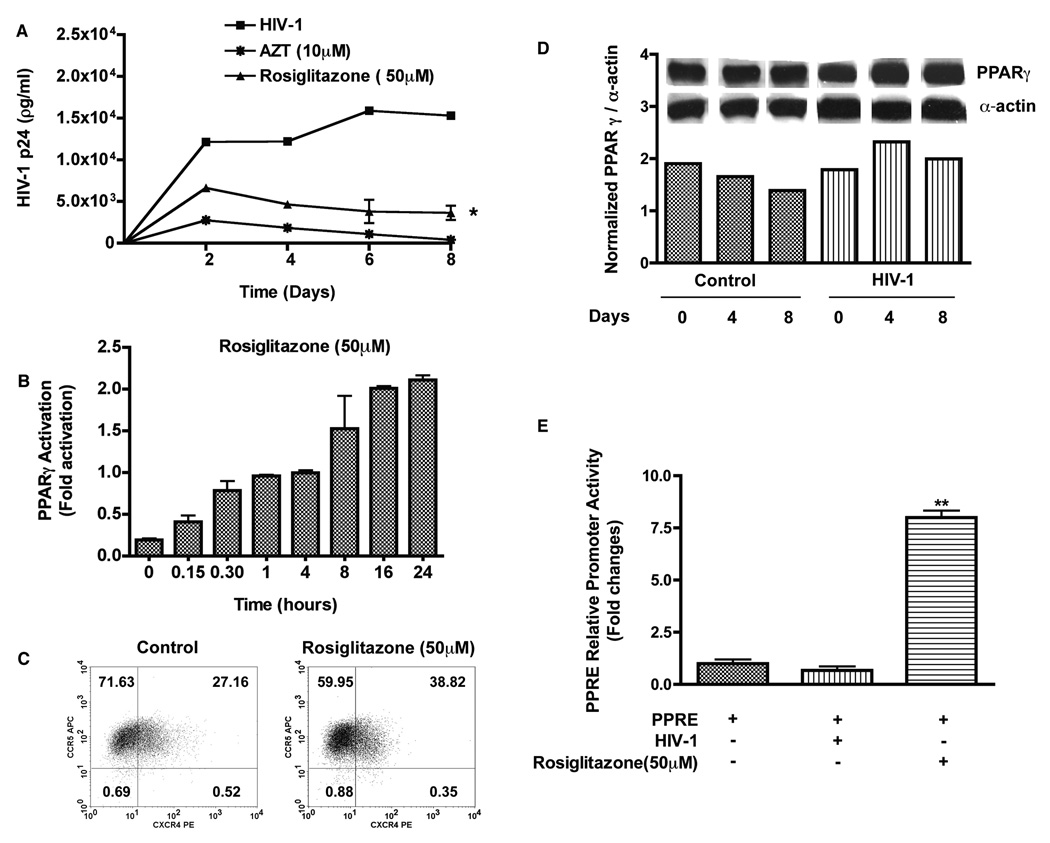
Figure 1. Rosiglitazone activates PPARγ and suppresses HIV-1 replication in MDM.
MDM were pre-treated with rosiglitazone (50 µM) for 18 hrs. Cells were infected with macrophage-tropic HIV-1ADA (at a multiplicity of infection of 0.1) for 12–18 hrs following which cultures were washed to remove any residual virus and replenished with fresh media. Half media exchange was performed every 2–3 days. (A) Levels of HIV-1 p24 antigen in culture supernatants. (B) Macrophages were treated with rosiglitazone (50 µM) for 0–24 hrs and the levels of PPARγ activity was measured by the TransAM PPARγ Kit. Results are expressed as fold changes in activation compared to control. (C) Representative flow cytometer scatter plots depicting CXCR4 and CCR5 expression in macrophages. (D) Expressions of PPARγ protein in control and HIV-1 infected human macrophages. (E) MDM were transfected with 1µg of PPRE luciferase reporter plasmid. Cells were harvested 24 hrs following infection or after treatment with rosiglitazone (50 µM) and luciferase activity of cell lysates was determined. Results are expressed as fold changes compared to basal luciferase activity. Data shown are mean ± SD of three independent experiments performed with three different MDM donors. *; P< 0.05 (PPARγ ligand treated vs. untreated HIV-1 infected MDM, **; P< 0.05 (Rosiglitazone treated vs. HIV-1 infected MDM.

