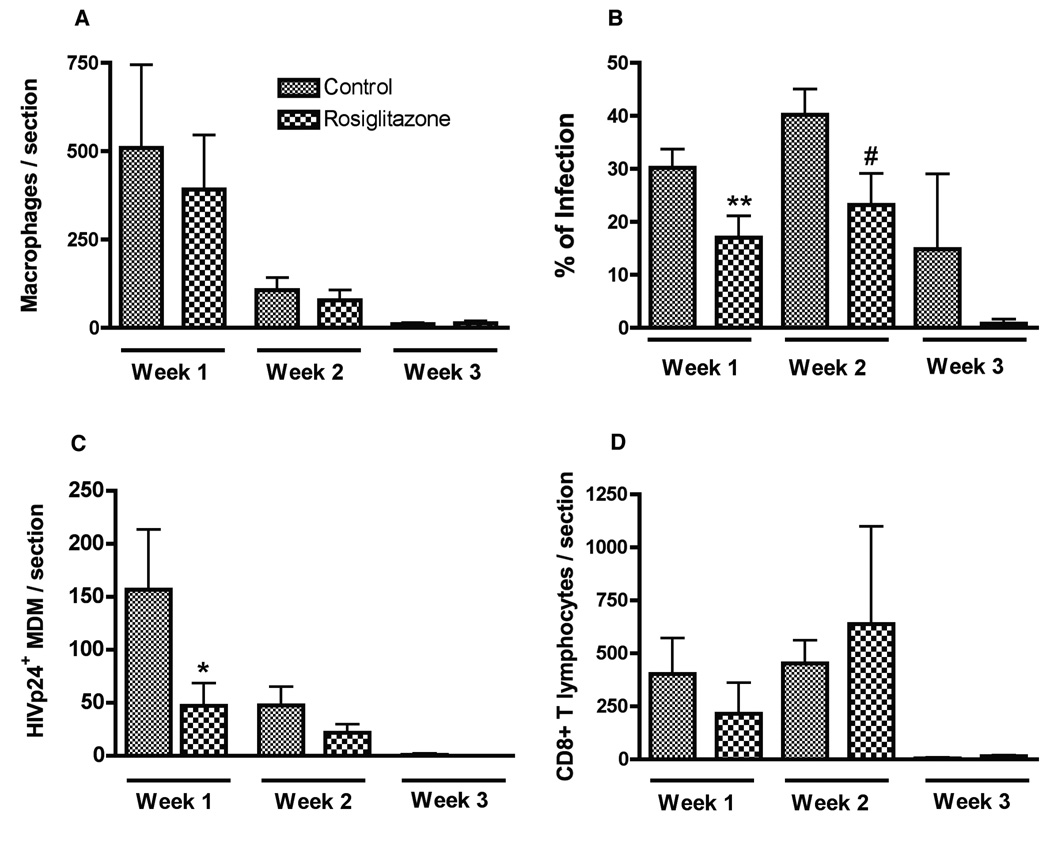
Figure 5. PPARγ ligand suppresses HIV-1 replication in brain macrophages in hu-PBL-NOD rosiglitazone /SCID HIVE mice.
Serial brains sections were immunostained for human macrophages (CD68+), HIV-1 p24+ MDM (viral antigen), and human lymphocytes (CD8+). Coronal brain sections were cut first to identify the injection site containing MDM. Serial 5-µm-thick sections of the brain (30 to 100) covering the entire area of human MDM injection were cut for each mouse brain and matched 3 to 7 slides (10 sections apart) were analyzed. Mean numbers of stained cells per section within the injected hemisphere were calculated for each mouse (3–7 sections/mouse), and a total of 5–8 mice per group were examined. Number of CD68+ MDM (A), percentage of infected MDM (B), number of infected MDM (C), and CD8+ lymphocytes (D) were counted in brain tissue section of control and rosiglitazone-fed hu-PBL-NOD/SCID HIVE mice. Rosiglitazone-fed mice demonstrated fewer HIV-1 p24+ MDM as compared to controls at week 1 (C). PPARγ treatment significantly decreased percentage of HIV-1 p24+ MDM rosiglitazone-fed mice at weeks 1 and 2 (B). Value represents mean ± SD per 5-µm section (n=5–8 mice/group). *,**, #; P< 0.05 (rosiglitazone vs. control).

