FIG. 1.

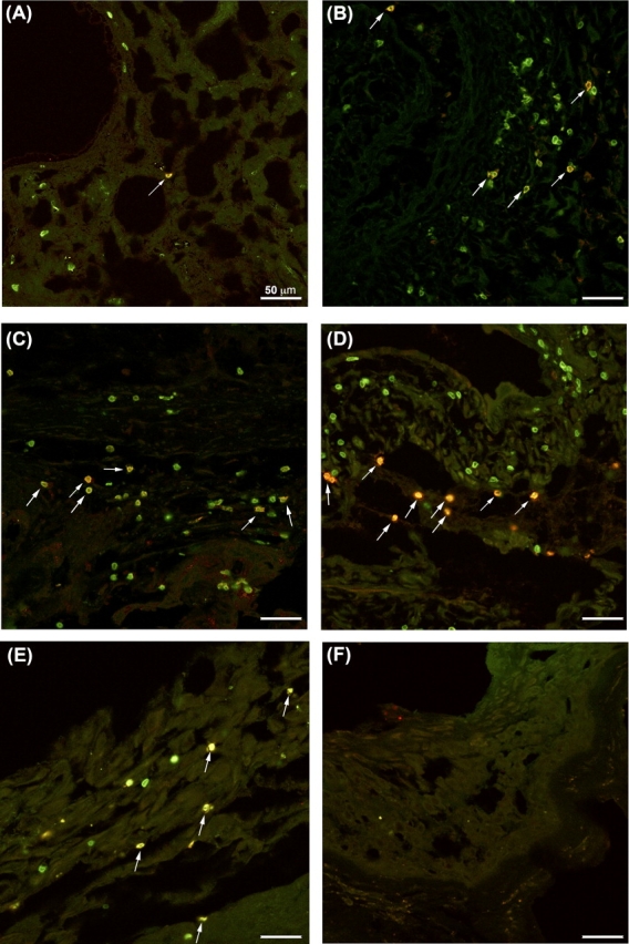
PDCD1 expression in endometrial and decidual tissues. Double-immunofluorescent immunohistochemistry of normal secretory endometrial tissues (A), first-trimester decidua (B), term basal plate (C), and term extraplacental membranes (D, E) with antibodies against CD3 (green) and PDCD1 (red). A–E) Photomicrographs are representative images of CD3 and PDCD1 immunolabeling for each group of tissues. F) Representative isotype control image from extraplacental membranes. Arrows indicate double-positive cells. Bars = 50 μm.
