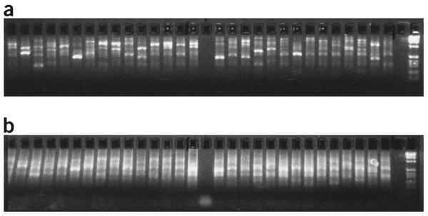
Figure 3.
Distinct banding patterns of DOP-PCR and amino-linking PCR products using long-insert genomic clones as templates. Note the much brighter smears—due to the increased DNA concentration—obtained after the secondary amino-linking PCR (b) compared with primary DOP-PCR (a). Negative controls for both PCRs are running in lanes 16 (a) and 16 (b).

