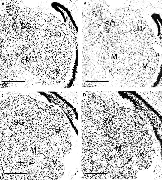
Figure 1.
Nissl stained coronal sections through the MGB from a P15 pallid bat pup. Arrows point to fiber tracts that separate MGBv and MGBm. Scale bars = 250 μm. A, B, C and D show sections cut at 15%, 25%, 50% and 75% from the caudal end of the MGB, respectively. D - dorsal division, M - medial division, V - ventral division, SG - suprageniculate nucleus.

