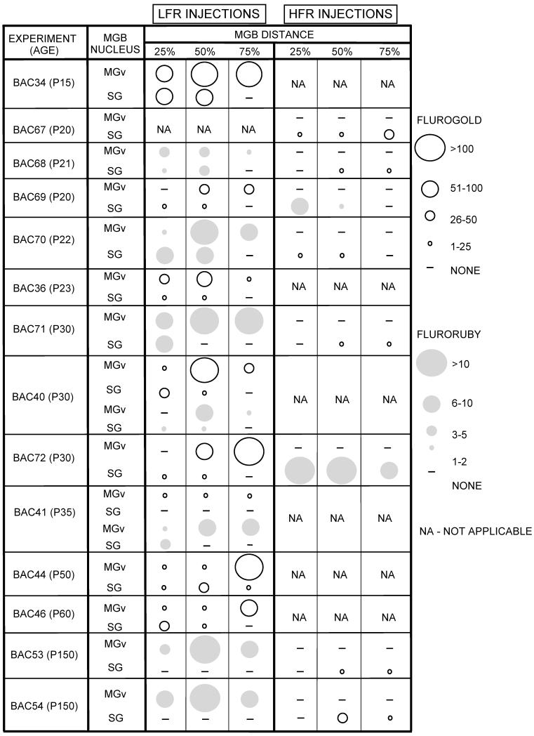
Table 1.
Summary of 14 experiments performed in this study. The number of labeled SG and MGBv neurons in three different caudorostral sections is shown schematically by the size of the circle. Light circles - FG. Gray circles - FR. Note that FG labeled more cells than FR. NA - not applicable (injection not made)

|
