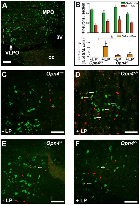
Figure 4. Effects of a 1-h light (L) pulse on c-Fos immunoreactivity in the VLPO.
(A) “Sleep-active neurons” of the VLPO identified by ISH contain galanin mRNA (labeled green). (B) Top: histograms represent the number of VLPO neurons expressing galanin mRNA or c-FOS protein per section (mean±SEM). Bottom: VLPO costained (galanin+c-Fos) neurons expressed as a percentage of the total number of galanin mRNA-positive neurons (mean±SEM). A two-way ANOVA (with factors light pulse and genotype) revealed that the L pulse induced an increase in VLPO costained cells in Opn4+/+, but not in Opn4−/− mice (Two-way ANOVA, L-pulse effect: p<0,05; L pulse×genotype interaction: a star indicates p<0.05; the asterisk [*] indicates post hoc Fisher PLSD: p<0.05). The light-induced immunoreactivity in VLPO in wild-type animals is specific to galaninergic neurons and of a large magnitude; however, the proportion of c-Fos-stained galanin neurons is low (e.g., Opn4+/+: +L pulse: 9% of total number of galanin mRNA-containing cells). (C) In the absence of a L pulse (control condition at ZT16), c-Fos immunoreactivity was found in nongalanin mRNA-containing neurons in the VLPO of Opn4+/+. (D) The L-pulse-induced c-Fos expression (labeled red) in some of the galanin mRNA-positive (green) neurons of the VLPO in Opn4+/+ (indicated by arrows). (E and F) In Opn4−/− mice, the same low number of galanin mRNA-positive neurons express c-Fos in both conditions, without (E) or with (F) light pulse. 3v, third ventricle; LP, L pulse administered during the habitual dark period (ZT15–16); MPO, medial preoptic area; oc, optic chiasm. Scale bars in (A) and (C–F) indicate 100 µm.

