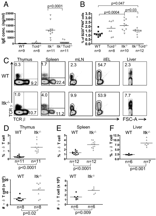
Fig. 1.
γδ T cells in Itk−/− mice are responsible for the spontaneous elevation in serum IgE levels. (A) Serum obtained from WT, Tcrd−/−, Itk−/−, and Itk−/−Tcrd−/− mice were analyzed for IgE by ELISA. (B) Splenocytes from WT, Tcrd−/−, Itk−/−, and Itk−/−Tcrd−/− mice were stained with α-B220 Ab and PNA to identify germinal center B cells. Differences between WT and Tcrd−/− (P = 0.14) and between Itk−/− and Itk−/−Tcrd−/− (P = 0.54) were not statistically significant. (C) Cells were prepared from thymus, spleen, mesenteric lymph nodes (mLN), intestinal epithelium (iIELs), and liver and were stained with anti-TCRδ and anti-TCRβ Abs. (D–F) Percentages and absolute numbers of γδ T cells were compiled for the thymus (D) and spleen (E); percentages only were compiled for liver (F).

