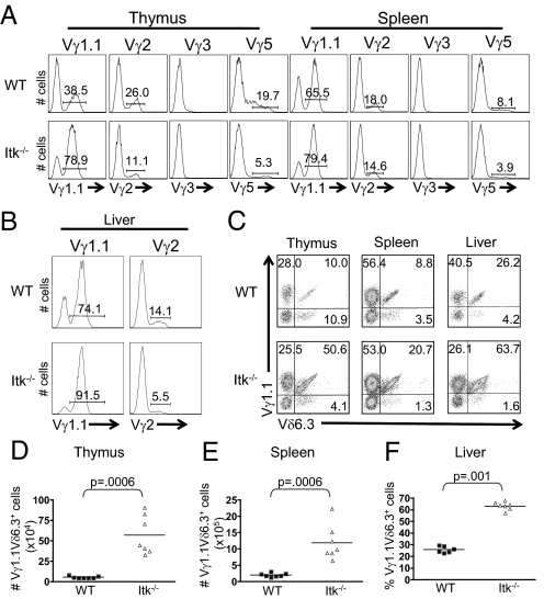
Fig. 2.
Itk−/− mice have an increased population of Vγ1.1Vδ6.3+ γδ T cells. (A) Thymocytes and splenocytes were stained with antibodies to TCRδ and either Vγ1.1, Vγ2, Vγ3, and Vγ5. Histograms show Vγ staining on gated TCRδ+ cells. (B) Lymphocytes were isolated from the liver and stained with antibodies to TCRδ and Vγ1.1 or Vγ2. Histograms show Vγ staining on gated TCRδ+ cells. (C) Thymocytes, splenocytes, and liver cells were stained with antibodies to TCRδ, Vγ1.1 and Vδ6.3. Dot plots show Vγ1.1 vs. Vδ6.3 staining on gated TCRδ+ T cells. (D) Absolute numbers of Vγ1.1Vδ6.3+ γδ T cells in the thymus and spleen and percentages of Vγ1.1Vδ6.3+ γδ T cells in the liver were compiled.

