Fig. 1.
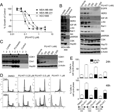
PU-H71 inhibits cell proliferation and blocks TNBC cells in G2-M. (A) Representative TNBC cells were incubated with increasing concentrations of PU-H71 and growth over 72 h was assessed. y-axis values below 0% represent cell death of the starting population. (B and C, Left) Hsp90-containing protein complexes isolated through chemical precipitation with beads having attached PU-H71 (PU-beads) or an Hsp90-inert molecule (control) were analyzed by western blot. Lysate, endogenous protein content; 1, MDA-MB-468; 2, MDA-MB-231; and 3, HCC-1806 cells. (Right) MDA-MB-468 cells were treated for 24 h with indicated concentrations of PU-H71, and protein extracts were analyzed by western blot. (D) MDA-MB-468 cells were treated for 24 h (Upper) or 48 h (Lower) with vehicle or with the indicated concentrations of PU-H71. DNA content was analyzed by propidium iodide staining and flow cytometry. (E) TNBC cells were treated for 24 h (Upper) or 48 h (Lower) with vehicle or PU-H71 (1 μM). The fraction of cells in G2-M and subG1 was analyzed by flow cytometry, quantified in FlowJo, and data were graphed.

