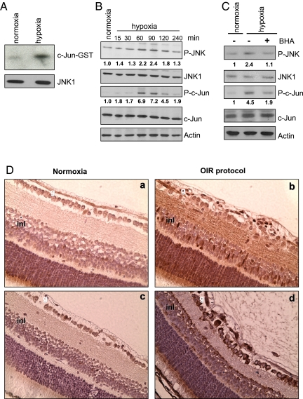
Fig. 2.
Hypoxia induces JNK activation. RAW264.7 macrophages were cultured either normoxia (Po2 = 21%) or hypoxia (Po2 = 0.5%). (A) Cell lysates were obtained after 1 h under normoxia or hypoxia, and JNK activity was measured by immunocomplex kinase assay using GST-c-Jun (1–79) as a substrate. (B) Amounts of phosphorylated (P) JNK and c-Jun were analyzed by immunobloting at different times after initiation of hypoxia. Numbers below P-JNK and P-c-Jun blots represent relative values normalized to total JNK and c-Jun amounts. (C) Cells were preincubated without or with BHA (10 μM) for 1 h, cultured under normoxia or hypoxia for 1 h, and analyzed as in B. (D) Retinas from P15 mice either under normoxia or subjected to OIR were stained with antibodies to phospho-c-Jun (a and b) or VEGF (c and d). g, ganglionar cell layer; inl, inner nuclear layer.

