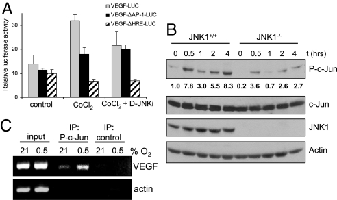
Fig. 4.
JNK1 regulates VEGF transcriptional activity. (A) RAW264.7 macrophages were transfected with pVEGF-Luc, pVEGFΔAP−1-Luc, or pVEGFΔHRE-Luc reporters. After 24 h, cells were treated with the hypoxia mimetic CoCl2 (200 μM). Some of the cells were pretreated with D-JNKi (1 μM) for 1 h. Results represent mean ± SEM luciferase activity relative to the internal control pRL-TK, in three separate experiments performed in triplicate. (B) Jnk1+/+ and Jnk1−/− BMDM were cultured under normoxia (Po2 = 21%) or hypoxia (Po2 = 0.5%). At different time points, cell lysates were prepared and analyzed for phosphorylated (P) or total amounts of the indicated proteins by immunoblotting. Numbers below P-c-Jun blot represent relative values normalized to total c-Jun.(C) ChIP was performed with anti-phospho-S63-c-Jun or a control antibody by using fixed and sheared chromatin isolated from RAW264.7 mouse macrophages cultured under normoxia or hypoxia for 1 h. The VEGF promoter fragment, which contains an AP-1 site at −1093/−1086 bp, was detected by PCR. The actin promoter served as a control.

