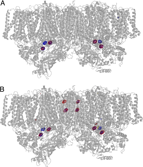
Fig. 2.
Difference Fourier maps and anomalous Fourier maps of PSII with Br− or I− substitution, superimposed on the structure of PSII dimer. (A) The difference Fourier map of PSII with Br− substitution minus Cl−-PSII and the anomalous Fourier map of PSII with Br− substitution measured at 0.9 Å were superimposed on the PSII dimer structure at 3.0 Å resolution (17) at a view perpendicular to the normal of the membrane plane. Red indicates the difference Fourier map contoured at σ = 6.0; blue: indicates the anomalous Fourier map contoured at σ = 6.0. (B) The difference Fourier map of PSII with I− substitution minus Cl−-PSII and the anomalous Fourier map of PSII with I− substitution measured at 1.0 Å superimposed on the PSII dimer structure at 3.0 Å resolution. Red indicates the difference Fourier map contoured at σ = 6.0; blue indicates the anomalous Fourier map contoured at σ = 5.0.

