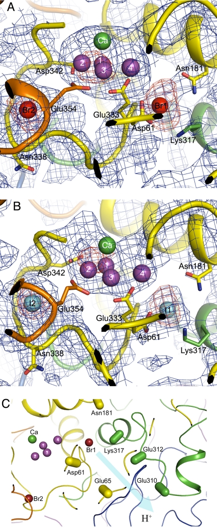
Fig. 3.
Location of the 2 anion-binding sites in PSII. (A) Composite omit Fo-Fc map (blue) and anomalous map (red) of PSII with Br− substitution contoured at σ = 1.0 and 4.0, respectively, superimposed on the structure of the Mn4Ca cluster and its surrounding regions. The structure of PSII with Br−substitution was shown by the molecular replacement method with the structure of the PDB code 2AXT as the search model. Color codes for the residues are as follows: yellow, D1; green, D2; orange, CP43; purple, Mn atoms. (B) Composite omit Fo-Fc map (blue) and anomalous map (red) of I−-substituted PSII contoured at σ = 1.0 and 4.0, respectively, superimposed on the structure of the Mn4Ca cluster and its surrounding regions. The structure of PSII with I− substitution was shown by the molecular replacement method. The color codes for the residues are the same as in A. (C) Location of Br1 relative to the proton exit channel proposed in (16, 27–29). The residues of PsbO are drawn in blue, and the residues of other subunits are in the same colors as in A. The directions of atoms from Cα to Cβ in the residues are indicated by capsule-shaped objects.

