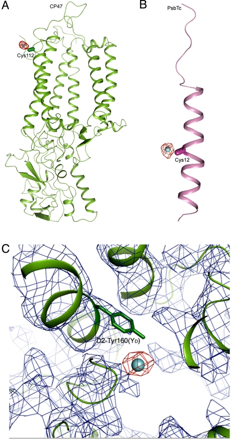
Fig. 4.
Additional binding sites of I− in I−-substituted PSII. (A) Fo-Fc omit map of PSII with I− substitution (red) contoured at σ = 3.0, superimposed on the structure of CP47 (green) obtained by the molecular replacement method, showing the association of I− with CP47-Cys-112. (B) Fo-Fc omit map of PSII with I− substitution (red) contoured at σ = 3.0, superimposed on the structure of PsbTc (pink), showing the association of I− with PsbTc-Cys-12. The capsule-shaped objects show the direction of atoms from Cα to Cβ in the 2 Cys residues. (C) 2Fo-Fc map (blue, σ = 1.0) and omit Fo-Fc map (red, σ = 2.0) of PSII with I− substitution around the region of D2-Tyr-160(YD), together with the structure of D2 (green) in this region.

