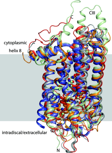
Fig. 1.
Structural superpositioning diagram of high-resolution crystal structures of bovine rhodopsin (red), squid rhodopsin (wheat), mutant β1-adrenergic receptor (light blue), mutant β2-adrenergic receptor (navy blue), and bovine opsin (gray) demonstrating a high level of overall structural similarity. Also shown are those water molecules that colocalize in the transmembrane helices.

