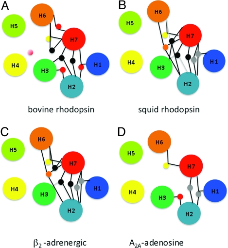
Fig. 3.
Conservation of hydrogen-bonding networks. Colocalized waters present in the crystal structures of bovine rhodopsin (A), squid rhodopsin (B), mutant β2-adrenergic receptor (C), and A2A-adenosine receptor (D) make contact with highly conserved amino acid residues. The lines connecting waters and helices indicate putative hydrogen bonds. For water clusters, yellow circles indicate conserved networks shared by all receptors, gray circles indicate interactions shared among squid rhodopsin, mutant β2-adrenergic, and A2A-adrenergic receptor, black circles indicate conserved networks shared among bovine rhodopsin, squid rhodopsin, and mutant β2-adrenergic receptor, red circles indicate conserved networks shared between bovine rhodopsin and mutant A2A-adenosine receptor, and finally, orange circles indicate conserved networks shared between squid rhodopsin and mutant β2-adrenergic receptor.

