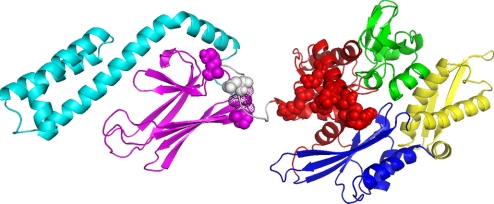
Fig. 3.
Mutagenesis and structure. The residues in E. coli DnaK, which when mutated affect the NBD–SBD interdomain communication, are represented as spheres in the colors of the subdomains to which they belong. These residues are, on the NBD: Y145A, N147A, and D148A (48); P143G and R151A (49); K155D and R167D (36). On the linker one has D393A (36). On the SBD they are K414I (31) and P419 (32, 33). Residues 417 and 418 SBD that show significant line broadening in the peptide-free form but not in the peptide bound form of the isolated SBD (11) are shown as dot surfaces.

