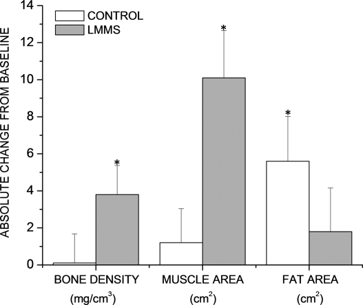
FIG. 7.
As measured by CT scans in the lumbar region of the spine, a group of young osteopenic women subject to LMMS for 12 mo (n = 24; gray bars ± SE) increased both BMD (p = 0.025 relative to baseline; mg/cm3) and muscle area (p < 0.001; cm2), changes that were paralleled by a nonsignificant increase in visceral fat formation (p = 0.45; cm2). Conversely, women in the CON group (n = 24; white bars ± SE), while failing to increase either BMD (p = 0.93) or muscle area (p = 0.43), realized a significant increase in visceral fat formation (p = 0.03). *Changes that are significantly different from baseline.

