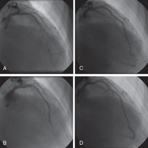
Figure 1).
Case 1. Angiography (right anterior oblique view) of the left anterior descending artery. A Initial angiogram demonstrating mid to distal vessel narrowing, no dissection plane identified. B Following percutaneous coronary intervention with bare metal stents, there is an excellent mid-vessel appearance with residual narrowing of the distal vessel. Thrombolysis in myocardial infarction 3 flow was demonstrated distally. C Second admission with chest pain, extension of dissection clearly evident in distal left anterior descending artery. The patient underwent further stenting with drug-eluting stents. D Repeat angiography demonstrated patent left anterior descending stents, with minor distal disease (unstented)

