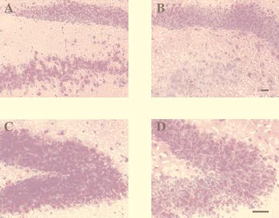
Figure 3.
(A) PAS staining of Sandhoff brain sections in the hippocampus (dentate gyrus). (A and C) Untreated brain. (B and D) NB-DNJ-treated brain. The images shown are representative of at least three animals per group. (Bar = 50 μm.) The reduction in PAS staining in NB-DNJ-treated mouse brains also was observed in other storage regions of the brain (data not shown). The data are from female mice, and comparable data were obtained from males (data not shown).

