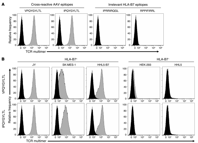
Figure 1. Antigen and HLA specificities of TCR multimer staining.
(A) To demonstrate antigen specificity, HLA-B*0702+ JY cells were pulsed with 5 μg/ml of either cross-reactive capsid epitopes derived from AAV2 (VPQYGYLTL) or AAV8 (IPQYGYLTL) or irrelevant HLA-B*0702–restricted epitopes derived from HIV (IPRRIRQGL) or EBV (RPPIFIRRL). After 2 hours of incubation, cells were stained with TCR multimer for flow cytometry. Histograms depict unpulsed cells (black) and peptide-pulsed cells (gray). (B) To demonstrate HLA specificity, either HLA-matched (JY, SK-MES-1, and HHL5-B7) or HLA-mismatched (HEK-293 and HHL5) cells were pulsed with 5 μg/ml of indicated AAV capsid peptide, then stained with TCR multimer. Histograms depict unpulsed cells (black) and peptide-pulsed cells (gray).

