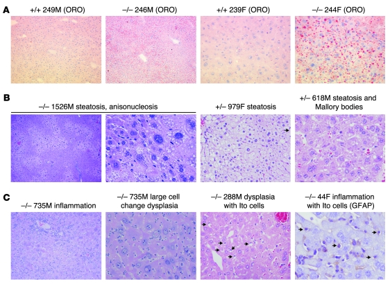
Figure 2. NAFLD and NASH with dysplasia in 10- to 12-week-old Taldo1–/– mice.
(A) Detection of lipid droplets with oil-red-O (ORO) staining in frozen liver sections of Taldo1–/– and Taldo1+/+ (+/+) littermates. Original magnification, left to right: ×100, ×100, ×400, ×400. (B) Centrolobular, zonal, or diffuse steatosis and Mallory bodies in Taldo1–/– and Taldo1+/– (+/–) mice. Original magnification, left to right: ×40, ×400, ×400, ×400. (C) Steatosis, inflammation, liver cell dysplasia with large cell change, and expansion of fat-storing hepatic stellate or Ito cells (arrows). Ito cells were also identified by expression of glial fibrillary acidic protein (GFAP). Original magnification, left to right: ×100, ×400, ×400, ×600.

