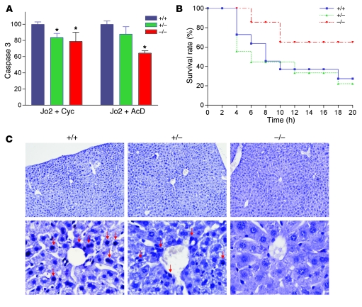
Figure 7. Resistance of Taldo1–/– hepatocytes to Fas-induced apoptosis.
(A) Fas-induced apoptosis of hepatocytes in vitro by activation of caspase 3. Hepatocytes were incubated as described in Methods with anti-Fas antibody (Jo2, 0.5 μg/ml) and 50 μg/ml cycloheximide (Cyc) or 50 ng/ml actinomycin D (AcD). Mean ± SEM values from 4 litters per independent experiment are expressed relative to hepatocytes from Taldo1+/+ littermates, set as 100%. *P < 0.05 versus Taldo1+/+. (B) Survival of 8- to 10-week-old Taldo1+/+ (n = 22), Taldo1+/– (n = 15), and Taldo1–/– littermate mice (n = 14) following i.p. injection with Jo2 antibody (10 μg/30 g body weight). Log-rank test showed increased survival of Taldo1–/– mice compared with Taldo1+/+ mice (P < 0.0001). No significant difference was observed between the Taldo1+/– and Taldo1+/+ groups. (C) TUNEL staining of liver sections 4 hours after i.p. injection with Jo2 antibody. Condensed and fragmented apoptotic nuclei (red arrows) in Taldo1+/+ and Taldo1+/– livers were visualized by staining with Vector Black substrate. Sections were counterstained with hematoxylin. Original magnification, ×100 (top row); ×400 (bottom row).

