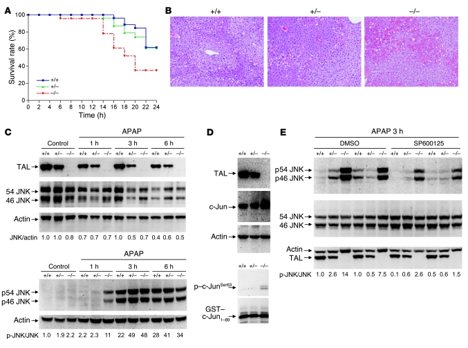
Figure 8. Increased susceptibility of Taldo1–/– mice to liver failure induced by APAP.
(A) Survival of Taldo1+/+ (n = 26), Taldo1+/– (n = 28), and Taldo1–/– littermates (n = 23) injected with 800 mg/kg APAP. Log-rank test showed reduced survival of Taldo1–/– compared with Taldo1+/+ mice (P = 0.027). No significant difference was observed between the Taldo1+/– and Taldo1+/+ groups. (B) H&E-stained liver sections obtained 6 hours after APAP injection. Hemorrhagic necrosis, characterized by hepatocyte vacuolization and extravasation of erythrocytes, was enhanced in Taldo1–/– liver. Original magnification, ×100. (C) Western blot detection of 46- and 54-kDa JNK and their state of phosphorylation (p46 and p54 JNK) in APAP-injected and untreated control mice. Numbers below blots show p-JNK/JNK levels, which were determined relative to actin and normalized to untreated Taldo1+/+ protein lysates, set as 1.0. (D) Assessment of JNK activity by in vitro phosphorylation of GST–c-Jun1–89 fusion protein in APAP-treated liver. TAL, c-Jun, and actin levels were detected by Western blot of liver cell lysates. In vitro phosphorylation of GST–c-Jun was detected by Western blot analysis using anti–phospho–c-JunSer63 antibody. (E) Effect of SP600125 on APAP-induced activation of JNK. Littermate 12-week-old mice were pretreated with SP600125 or DMSO control as described in Methods 1 hour prior to APAP exposure. Numbers below blots show p-JNK/JNK levels 3 hours after APAP treatment; values were determined relative to actin and normalized to untreated Taldo1+/+ protein lysates, set as 1.0.

