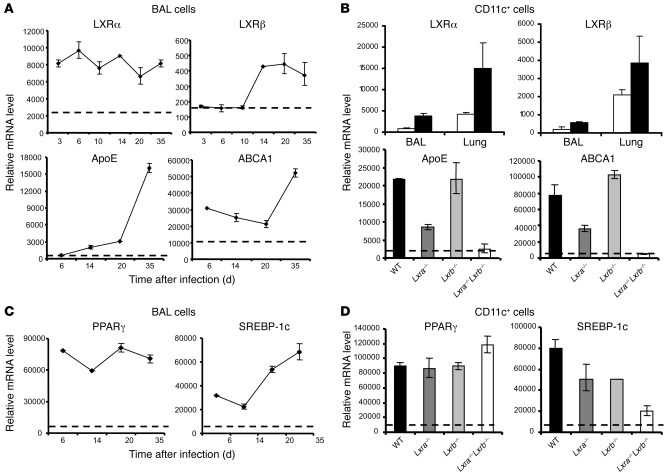
Figure 2. Expression of LXRs and LXR-dependent genes during M. tuberculosis infection.
(A and C) RT-qPCR analysis of expression levels of LXRα and LXRβ isoforms, LXR-dependent target genes, and PPARγ and SREBP-1c within BAL cells isolated at the indicated times after infection. Dotted lines denote mRNA levels obtained from background-matched naive BAL cells. Mice were tested individually (n = 4–5). (B and D) After 3 weeks of infection, CD11c+ cells were isolated from the BAL, and lung tissue digests and relative mRNA levels of the indicated parameters were determined by RT-qPCR. (B, top) Infected (black bars) CD11c+ BAL and lung cells or naive controls (white bars) from WT mice. (B, bottom, and D) Transcriptional analysis of CD11c+ BAL cells isolated from infected WT, Lxra–/–, Lxrb–/–, and Lxra–/–Lxrb–/– mice (n = 5). Dotted lines denote mRNA levels obtained from background-matched naive CD11c+ BAL cells.

