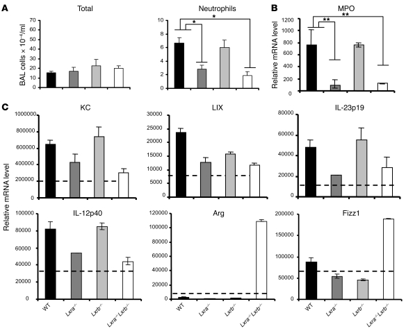
Figure 5. LXR-dependent regulation of the innate immune response after i.t. challenge with M. tuberculosis.
WT, Lxra–/–, Lxrb–/–, and Lxra–/–Lxrb–/– mice were infected i.t. with luminescent M. tuberculosis (1 × 10⁴ CFU) and sacrificed 7 days after infection. (A) Differential cell infiltration in the BAL. Shown are the absolute numbers of total cells and neutrophils. Data in A are mean ± SEM (n = 5). (B) Relative mRNA levels of MPO from total lung tissue samples, as determined by RT-qPCR (n = 5). (C) CD11c+ cells were isolated 7 days after infection from lung tissue digests, and relative mRNA levels were determined by RT-qPCR. Dotted lines denote values obtained from CD11c+ cells of naive mice. Data are expressed as relative mRNA levels, normalized against reference housekeeping genes (n = 5), and are representative of 2 separate experiments. Arg, arginase. *P < 0.05, **P < 0.01.

