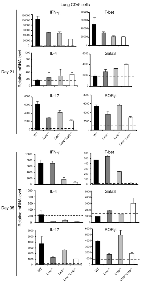
Figure 6. The role of LXRs in the modulation of T cell function during M. tuberculosis infection.
WT, Lxra–/–, Lxrb–/–, and Lxra–/–Lxrb–/– mice were infected i.t. with luminescent M. tuberculosis (1 × 10⁴ CFU). After 21 and 35 days of infection, CD4+ lung T cells were isolated, and the relative mRNA levels of Th1 (IFN-γ and T-bet), Th2 (IL-4 and Gata3), and Th17 (IL-17 and RORγt) markers were analyzed by RT-qPCR. Data are expressed as relative mRNA levels, normalized against reference housekeeping genes (n = 5). Results are representative of 2 separate experiments.

