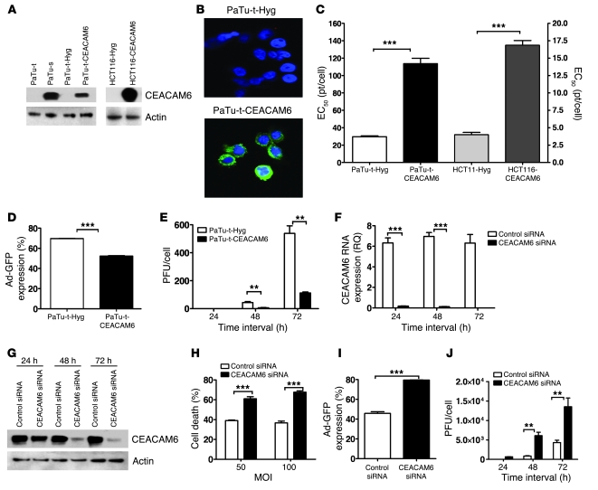
Figure 3. CEACAM6 functionally affects adenovirus infection.
(A) Expression of CEACAM6 protein in pancreatic cancer cell lines and engineered subclone cell lines as analyzed by Western blotting. (B) CEACAM6 expression in vector-transfected PaTu8988t (top) and CEACAM6-transfected PaTu8988t cell lines (bottom) by confocal microscopy, showing CEACAM6 expression on membrane and in cytoplasm. Blue (DAPI staining) indicates nuclei; green, CEACAM6; original magnification, ×600. (C) Sensitivity of CEACAM6-overexpressing cell lines and their counterparts to adenovirus as assayed by MTS. The EC50 values were increased 4-fold in CEACAM6-transfected PaTu8988t and HCT116 cells compared with their counterparts. (D) The infectivity of adenovirus Ad-GFP in stable clones of PaTu8988t-Hyg and PaTu8988t-CEACAM6 by FACS analysis at 48 hours after infection with Ad-CMV-GFP adenovirus. (E) Adenovirus replication in stable clones of PaTu8988t-Hyg and PaTu8988t-CEACAM6 (infected at an MOI of 100 pt/cell). (F) The expression of CEACAM6 as analyzed by qPCR in Suit-2 cell line after treatment with control siRNA and the CEACAM6-specific SMARTpool at various time points. (G) The expression of CEACAM6 protein by Western blotting after treatment with control siRNA, and the CEACAM6-specific SMARTpool at various time points. (H) Cell death of control siRNA– and CEACAM6-specific SMARTpool siRNA–pretreated Suit-2 cells after adenovirus infection at MOI of 50 and 100 pt/cell. (I) The infectivity of adenovirus Ad-GFP in control and CEACAM6-specific SMARTpool siRNA–pretreated Suit-2 cells by FACS at 48 hours after infection with Ad-CMV-GFP adenovirus. (J) Adenovirus replication in control and CEACAM6-specific SMARTpool siRNA–pretreated Suit-2 cells (infected at an MOI of 100 pt/cell) by 50% tissue culture infective dose (TCID50) assay. All experiments were repeated at least 3 times. **P < 0.01, ***P < 0.001.

