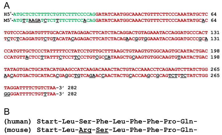
Fig. 4.
(A) Nucleotide sequence alignment of the human (top rows) and mouse (bottom rows) TSH splice variant. Green nucleotides are the putative intron-coded leader sequences. Red nucleotides represent exon 3 and exon 5 of the human and mouse TSH gene, respectively. Underlined black nucleotides show the difference of the mouse relative to the nucleotide in the corresponding human gene. (B) Comparison of amino acids in the putative leader sequence of the human and mouse TSH splice variant, indicating species differences at amino acid positions three and four.

