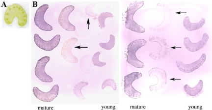
Figure 3.
(A) Scanned image of a cross-section of fresh celery petiole. (B) Examples of student-prepared tissue prints showing distribution of Rubisco on cross- sections of celery petioles. The print was produced by pressing the cut surface of celery petioles on a piece of nitrocellulose membrane, which was then incubated with anti-Rubisco primary antibody, followed by a secondary antibody conjugated to alkaline phosphatase. Purple color indicates the presence and location of Rubisco. Cross-sections shown are from young and mature celery petioles. Arrows indicate prints from celery petioles kept in water and under dim light for several days before prints were made, showing decreased Rubisco content.

