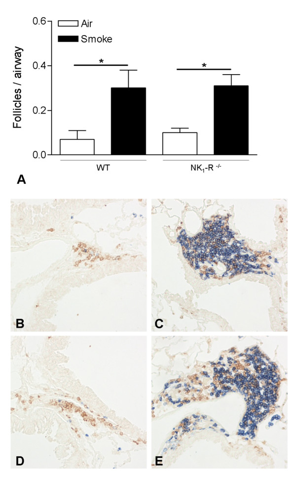
Figure 2.

Quantification of pulmonary lymphoid follicles upon chronic cigarette smoke exposure. Peribronchial lymphoid follicles in lung tissue of wild type and NK1-R-/- mice upon chronic (24 weeks) exposure to air or cigarette smoke (CS) (A). Results are expressed as means ± SEM. N = 8 animals per group (* p < 0.05). Photomicrographs of peribronchial lymphoid follicles in lung tissue of air- and CS-exposed wild type and NK1-R-/- mice at 24 weeks (chronic exposure; magnification ×200): (B) air-exposed wild type mice, (C) CS-exposed wild type mice, (D) air-exposed NK1-R-/- mice and (E) CS-exposed NK1-R-/- mice.
