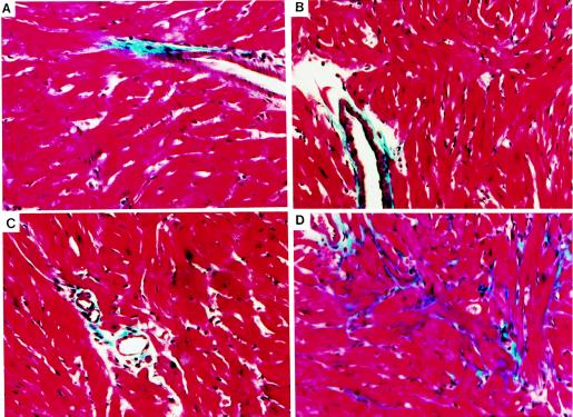
Figure 7.
Myocardial fibrosis in Gαq/β2ARM transgenic mice. Masson’s trichrome stain of myocardial section from mid-left ventricular free wall of 8-week-old NTG (A), Gαq (B), Gαq/β2ARL (C), and Gαq/β2ARM (D). The perivascular blue staining serves as a control for the stain in that it identifies vascular collagen. Gαq/β2ARM exhibits significant fibrosis not observed in other groups (representative of 4–6 individual hearts examined). (Magnification, ×200.)

