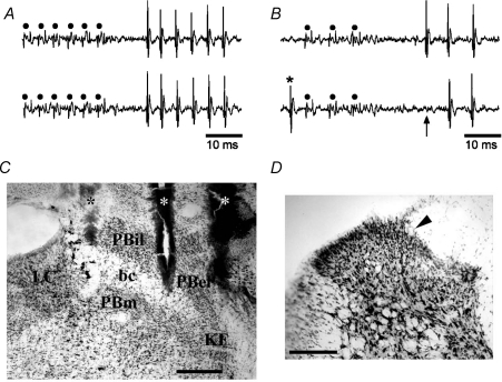
Figure 1. Identification of lamina I spinoparabrachial neurons in vivo.
A, pair of traces showing 1-for-1 following of a train of 6 antidromic electrical stimuli (45 μA, 2 ms, 250 Hz; dots) delivered from the middle stimulating electrode in the contralateral parabrachial nucleus. B, collision of the first antidromic impulse in a train of 3 (150 Hz, upper trace) when an orthodromic impulse (asterisk, lower trace) occurred within the critical interval. The arrow indicates the point at which the first antidromic response should have occurred. C, photomicrograph of a frozen section stained with thionin showing the tracks of the stimulating electrodes (asterisks) in the contralateral parabrachial nucleus. Bar is 0.4 mm. bc, brachium conjunctivum; KF, Kölliker–Fuse nucleus; LC, locus coeruleus; PBel, external lateral subnucleus of the parabrachial area; PBil, internal lateral nucleus of the parabrachial area; PBm, medial subnucleus of the parabrachial area. D, photomicrograph of the contralateral spinal dorsal horn at the level of the 3rd lumbar segment. An arrowhead marks the position of an electrolytic microlesion that was made at the recording site of the cell shown in A and B. Dorsal is up, lateral is left. Bar is 0.2 mm.

