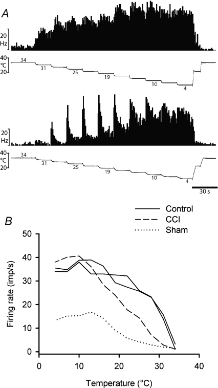
Figure 4. Stimulus encoding by lamina I spinoparabrachial COOL neurons.
A, peristimulus time histograms from two cooling-specific neurons showing their responses to graded intensity cooling stimuli. The upper pair of records was from an unoperated control animal and the lower pair was from an animal that had received a CCI. B, stimulus–response curves of all 4 cooling-specific neurons isolated in the current study.

