Fig. 1.
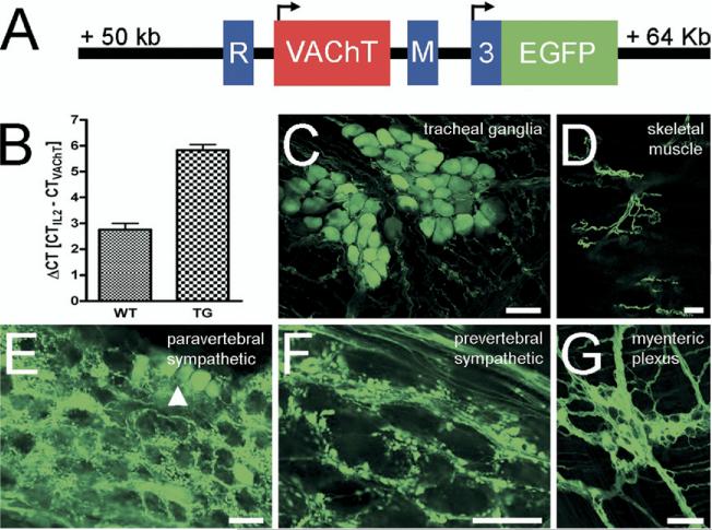
EGFP is expressed in a cholinergic-specific manner from the CGL in ChAT-EGFP transgenic mice. (A) Schematic representation of the core region from the CGL, as modified in the ChAT-EGFP BAC clone. The EGFP coding sequence (green color) is integrated into the first ChAT coding exon (no. 3, blue color). Two additional, 3-prime non-coding ChAT exons (R and M) flank a single VAChT-encoding exon (red color). Translation start sites for the two genes are marked by arrows. (B) Estimation of the VAChT, and hence BAC copy number in hemizygous transgenic mice by qPCR. In wild type (WT) mice, a ΔCT=2.75±0.25 (n=5) equals two VAChT copies per diploid genome. In hemizygous ChAT-EGFP transgenic mice, a ΔCT=5.83±0.23 (n=7) equals additional eight transgene copies. (C–G) Examples for EGFP fluorescence in native preparations from selected authentic cholinergic peripheral nerves. Parasympathetic tracheal ganglia (C), motor nerves in skeletal muscle (D), post-ganglionic cholinergic neurons (arrowhead) and pre-ganglionic nerve terminals in a paravertebral sympathetic ganglion (E), pre-ganglionic nerve terminals in a prevertebral sympathetic ganglion (F), and the myenteric plexus (G) display EGFP fluorescence. Scale bars=50 μm.

