Fig. 2.
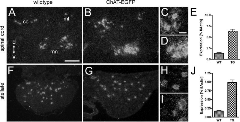
VAChT mRNA is up-regulated in adult ChAT-EGFP transgenic mice. Darkfield microphotographs of VAChT ISH signals (left three columns), and changes in VAChT mRNA expression levels determined by RT-PCR (right column) from wild type (WT) and ChAT-EGFP transgenic (TG) mice. (A) Transverse section through upper lumbar spinal cord. VAChT mRNA expressing neurons are around the central canal (cc), in the intermediolateral cell column (iml), and the ventral horn motor neuron area (mn). Scale bar=100 μm (also applying to B, F, and G). d=Dorsal, v=ventral. (B) Comparable section to (A) from a TG mouse, showing intensification of VAChT ISH signals as compared with WT. (C) Higher magnification of motor neuron area from (A). Scale bar=25 μm (also applying to D, H, and I). (D) Higher magnification of motor neuron area from (B). (E) VAChT mRNA is up-regulated fivefold in spinal cord of TG compared with WT mice. (F) SG from a WT mouse. Several neurons exhibit VAChT ISH signals. (G) SG from a ChAT-EGFP mouse. ISH signal intensities for VAChT are increased as compared with WT. (H, I) Higher magnification from (F) and (G), respectively. (J) VAChT mRNA is up-regulated fivefold in TG SG as compared with WT.

