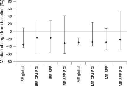
Figure 3.
Median percentage change in key magnetic resonance imaging (MRI) measures. MRI of the knee was performed, and measures of the initial rate of enhancement (IRE) and maximal enhancement (ME) made in the global region as well as in regions of interest (ROI) at the cartilage–pannus junction (CPJ) and the suprapatellar pouch (SSP). Values for the ME and IRE are calculated from the sum of the enhancing pixels in each region of interest, expressed as arbitrary units. Error bars represent 25th and 75th percentiles.

