Fig. 2.
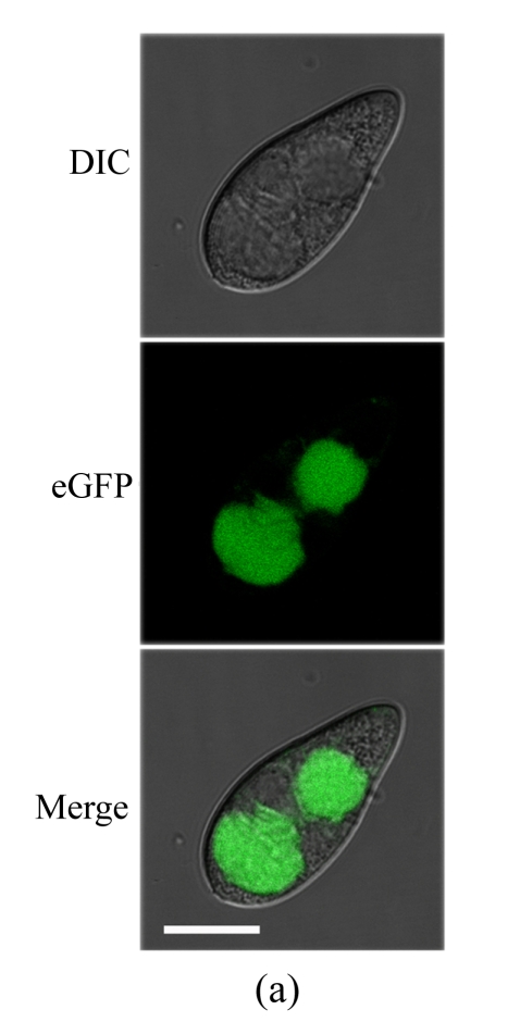
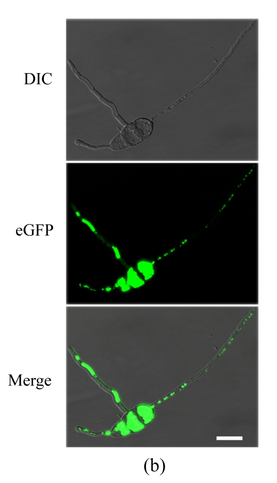
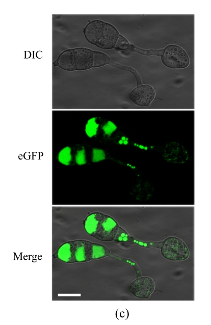
Cellular localization of MoFLP1 in M. oryzae. MoFLP1-GFP protein was detected in the vacuoles of M. oryzae strain NMG2
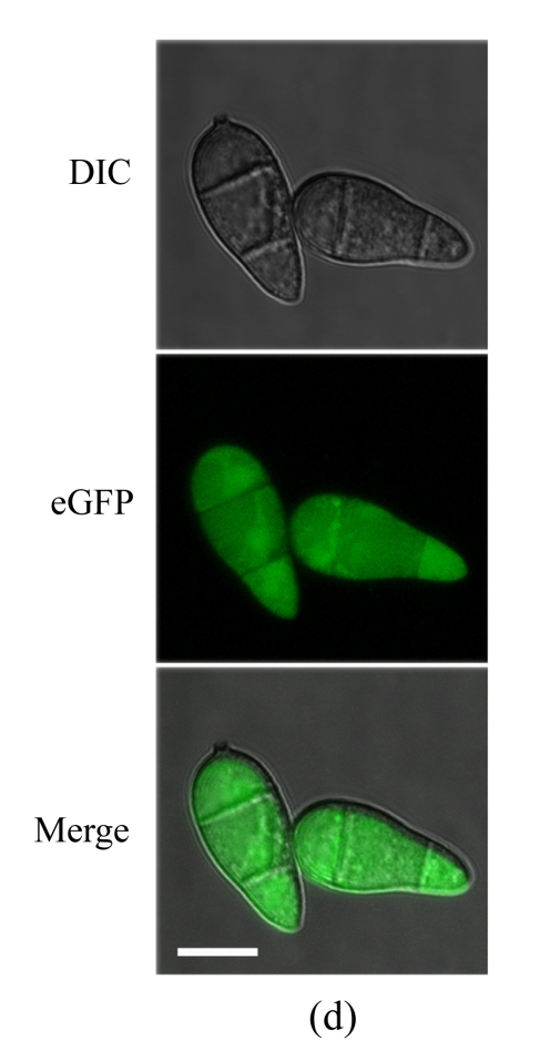
The conidia (a), germinated conidia (b) at 10 h post-incubation, and appressoria (c) at 6 h post-incubation of strain NMG2 were observed by inverted confocal laser scanning microscope. Representative bright-field [differential interference contrast (DIC), top], fluorescence (middle), and merged images (bottom) are shown. (d) The conidia of the Guy11 strain transformed with pGFP-ATG1 were observed by inverted confocal laser scanning microscope and ATG1-GFP appeared in the cytoplasm. Bar=10 μm


