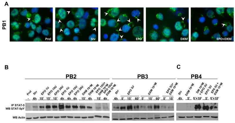
Figure 5. Levels of STAT-5 phosphorylation after exposure either to GF starvation, EPO and DXM, alone or in combination, of human proerythroblasts obtained from normal donors.
A) Immunofluorescence analyses with an anti-STAT-5 antibody of proerythroblasts obtained in HEMA culture (Prol) from a normal donor and treated as indicated in Figure 3B. Original magnification: 100×. Representative cells in which STAT-5 is localized on the plasma membrane are indicated by arrowheads. The microscopic images were acquired as described in the legend of Figure 3A. Quantification of the level of STAT-5 immunostaining in the nuclear area is presented in Table 1.
B) Levels of STAT-5-phosphorylation in proerythroblasts obtained in HEMA culture (Prol) from two normal donors (each panel a different donor), GF starved for 4 h (Stv) and then treated for 5′-4h either with EPO (1–30 U/mL), DXM (10−6 M) or the combination of both, as indicated. The specificity of the signal for the phosphorylated form of STAT-5 was increased by analyzing proteins that were immunoprecipitated from 50 μg of whole cell lysates with a STAT-5 antibody by western blot with an anti-STAT-5pY antibody. Equivalent amounts (10 μg) of pre-immunoprecipitated proteins were analyzed by western blot with an anti-Actin antibody, as quantitative control. Similar results were obtained in 10 additional experiments.
C) Levels of STAT-5-phosphorylation in proerythroblasts preincubated for 5′ either with DXM (10−6 M) or EPO (3 U/mL) and then exposed to EPO (3U/mL) or DXM (10−6 M) for 10′.

