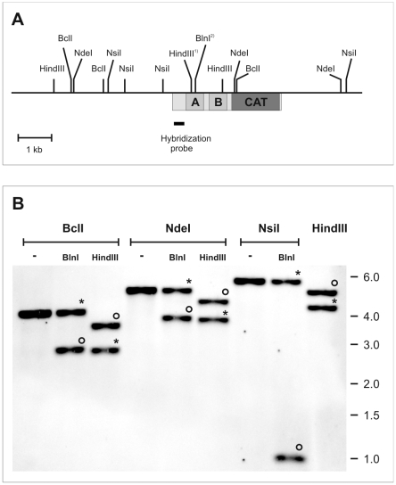
Figure 2. Restriction enzyme analysis of TbrPDEB2a and TbrPDEB2b.
Panel A: Restriction map of the TbrPDEB2 locus on chromosome 9. HindIII1): site present only in the converted allele TbrPDEB2b; BlnI2): site present only in non-converted allele TbrPDEB2a. Box underneath horizontal line: Open reading frame of TbrPDEB2. A,B: GAF domains, CAT: catalytic domain. Hybridization probe: detects both alleles, TbrPDEB2a and TbrPDEB2b; corresponds to nucleotides 158–518 of TbrPDEB2. Panel B: Southern blot analysis of singly or doubly digested genomic DNA establishes the presence of both alleles in the genome of strain Lister427. Circles: Fragments derived from allele TbrPDEB2a; asterisks: fragments derived from allele TbrPDEB2b; no tag: fragments common to both. Sizes of molecular weight markers are indicated to the right.

