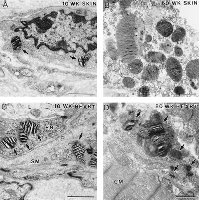
Figure 3.
EM analysis of skin and heart sections from α-Gal A −/0 mice of different ages. (A) Ten-week-old skin section showing a macrophage-like cell containing two distinct lipid inclusions (arrows). (B) In the skin section of 60-wk-old mice, the lipid inclusions increased in size and number. (C) Heart section from 10-wk-old mice showing typical lipid inclusions. An endothelial cell (EN) lining the lumen (L) of a blood vessel and a smooth muscle cell (SM) surrounding the blood vessel contain the characteristic lipid inclusions (arrows). (D) Heart section from 80-wk-old mice. A macrophage-like cell in the connective tissue had many typical lipid inclusions (arrows); some of them were very large in size. CM, cardiac muscle. (Bar = 1 μm.)

