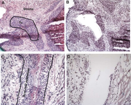
Fig. 1.
Microdissection of squamous cell carcinoma tumors and normal bronchial epithelium. Tissue sections were stained with hematoxylin and eosin. Neighboring sections stained only with hematoxylin were used for needle microdissection. Tumor (Tu) tissue (A, before and B, after needle micro-dissection) was specifically enriched and separated from surrounding stroma (C, before and D, after micro-dissection) just like bronchial epithelium was specifically enriched and separated from surrounding connective tissue and smooth muscle cells.

