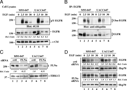
Figure 2.
Effect of EGF on ligand-mediated EGFR activation and down-regulation in highly metastatic human melanoma cell lines. A, M93-047 and UAC647 cells were fed serum-free medium for 3 h and then left alone or incubated with EGF (20 nm) for 2.5, 10, and 30 min. Cells were lysed and analyzed by Western blot with antibodies against phosphotyrosine (pY, clone 4G10), EGFR, or c-Cbl as a loading control. Densitometry analysis was carried out and normalized to c-Cbl. The EGFR signal in unstimulated cells (second panel, lanes 1 and 5) was arbitrarily given the value of 1.0. B, M93-047 and UACC647 cells were treated as in A. EGFR immunoprecipitates (IP) prepared from cell lysates were resolved by SDS-PAGE and then analyzed by Western blot with antibodies against ubiquitin, or EGFR. Ubn-EGFR, Ubiquitinated EGFR. C, M93-047 and UACC647 cells were transfected with either control (crtl) or FLNa siRNA for 72 h, lysed and then analyzed by Western blot with antibodies against FLNa or ERK1/2 as a loading control. Densitometry analysis was carried out and normalized to ERK. The FLNa signal in crtl siRNA-transfected cells was arbitrarily given the value of 1.0. D, Time course of EGF stimulation was carried out in cells transfected with control (crtl) or FLNa siRNA. Cell lysates were prepared and immunoblotted for total EGFR or Hsp70 as a loading control. Densitometry analysis was carried out and normalized to Hsp70. The EGFR signal in unstimulated cells was arbitrarily given the value of 1.0. Data shown in C and D are representative of two independent experiments, each performed in duplicate dishes.

